(01.03.06/01.01.2008*)


go back

Estrogen Made Easy(er)


Effects of Estrogen in Women
Dr. Timothy Bilash MD OBGYN
January 2006

www.DrTimDelivers.com
  • The Menopause Problem

    Menopause has become a bit like the weather: almost impossible to avoid, and everybody has an opinion about it. I fully expect "The Menopause Channel" to pop up on cable one of these days, forecasting daily with psychedelic maps and ever changing expert current forecasts.

    from www.everythingmenopause.com
    from www.healthsquare.com
    &
    BODY
    SOUL
    DrTimDelivers.com endeavors to snapshot the evidence that Estrogen, of any type, is not a devil to be feared, but a required medicinal to be carefully and knowledgeably prescribed. The following reviews many of the known health benefits of Estrogen; It's time to put the pieces together. (This review omits an extensive amount of information known about Progestin, an important co-factor affecting health and cancer.)


    Clinical Benefits of Estrogen (Easy Part)

    1. Bone, Joints and Skin
      1. Osteoporosis, which imobilizes more women than any other disease in
        advanced old age, is entirely prevented with Estrogen treatment. Estrogen
        makes bones strong, improving Calcium content and Vitamin D levels.
        Estrogen inhibits Osteoarthritis. 1
      2. Bone and Joint pain is associated with Estrogen deficiency, and reversed with
        Estrogen therapy. 2
      3. Skin looks younger and softer with Estrogen 3
        1. 30-50% of skin collagen depends on Estrogen, which prevents and
          repairs damaged skin. 3a
        2. Small blood vessels that nourish the skin, vaginal skin health and
          lubrication, requires Estrogen
        3. Low Estrogen impairs wound healing 4

    2. Brain Function and Sleep
      1. Improves sleep and integration of brain activity 12
      2. Improves memory 6 , inhibits Alzheimers onset 7
      3. Improves coordination 8
      4. Improves mood, stabilizes lability (loss of control, emotions) 8

    3. Heart & Vascular Disease
      1. Inhibits arterial plaques (arterial disease) caused by low Estrogen 10
      2. Maintains smooth muscle function of blood vessels (better blood flow and
        tissue perfusion) 11
      3. Lowers total Cholesterol, increases HDL 12
      4. Lowers Heart Disease and Mortality when started near menopause 13

    4. Mortality
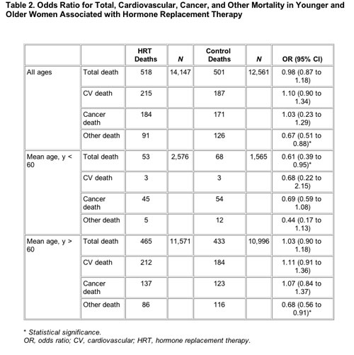
      1. Observational Studies (retrospective and prospective) indicate the same or lower
        mortality with Estrogen and Estrogen/Progestin therapy. WHI does not contradict these findings, (and in fact a further analysis of WHI now shows a decreased mortality of 30% between 50-60 years of age with either Estrogen or Estrogen+Progestin).(c) 14
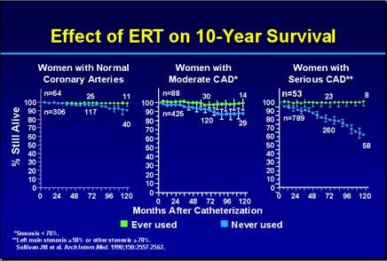
      2. Estrogen benefit for Mortality from Cardiac Disease is highly dependent on the
        severity of disease 15
      3. Estrogen treatment decreases mortality after Breast Cancer(a) 16,49

    5. Cancer
      1. Overweight women have a higher risk of breast cancer (the major risk factor) 17
      2. Apoptosis (programmed cell disintegration) plays an important role in
        preventing cancer, is modulated by steroid hormones 18
      3. Breast cancer risk is associated with an increase in Free Estradiol produced from the Tumor, not causing the Tumor (as with Menopausal transition, Obesity or Anovulation)(b) 19
      4. Breast cancer cell lines resistant to Estrogen deprivation undergo cell death
        when treated with menopausal concentrations of estradiol. The Estrogen is
        100% effective in killing the cancer cells. 46

    6. Hot Flashes
      1. Currently the least controversial benefit for Estrogen is treatment of hot flashes 20

    7. Sex Drive
      1. Libido and energy depend on Free Testosterone (converted from Estradiol) 21,51

    8. Obesity
      1. Estrogen reduces tummy fat at menopause 22
      2. Estrogen lowers blood sugar and prevents onset of diabetes 24

    9. Immune System
      1. Estrogen improves Immune function and Rheumatoid Arthritis 25

    10. Thrombosis concern note
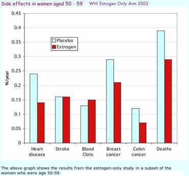
      1. Hemorrhagic Stroke (caused by bleeding, more severe, 10% of strokes) is
        reduced by Estrogen, but Thrombotic Stroke (ischemia caused by clotting,
        less severe, 90% of strokes) may be increased by Estrogen (the evidence is
        conflicting). 26
        This is mostly seen in the first year of use and for older women, with indications
        of no increase for total stroke in the younger age group 50-59. Stroke mortality
        is not affected in any age group.
        1. Estrogen actually increases the rate of clot lysis. 27
        2. Estrogen keeps brain tissue healthier, but may adversely affect brain tissue
          that is already damaged. Neural blood vessels 8 and nerve growth 6
          are adversely affected by a lack of Estrogen.
        3. Thrombotic risks may be related to increased levels of Free rather than
          Total Estrogen, and not in a simple way.
      2. Statins, Aspirin (COX-1) and certain tNSAIDS ("traditional" less COX selective) alter
        cardiovascular risk and the effect of estrogen. They appear to lower Thrombotic
        Stroke and Heart Attack rates, preventing initial adverse effects of hormone
        supplements in older, high-risk women. 30 Discontinuing Aspirin or NSAIDS,
        in contrast increases Stroke and MI. 48
      3. Age and vascular disease severity appears to increase the risk of Thrombotic
        Stroke and MI, and effects of estrogen and NSAIDS.

  • Latest News (Winter 2005)

    1. "Benefits outweigh Risks for women to start estrogen at menopause." 13
      The conclusions of the Women's Health Initiative (WHI) are being discredited. The WHI Hazard Rates for events were actually better when starting estrogen-progestin in the 50-59 year-old perimenopausal group (Stroke and Blood Clots was not a factor in this age group). Taking appropriately dosed Estrogen supplements does not cause cancer (although it may uncover it), and certainly does not increase mortality rates. It is important to consider early supplementation before the levels of Estradiol drop. Maintaining the right amounts and timing 21 of Estrogen, adjusting to the levels of other hormones is advised (not too little, not too much). Birth Defects and Cancer is actually more strongly associated with low rather than high blood levels of Total Estrogen. 28 The details of this are still being investigated.

    2. Statistical significance within the errors of the data should be demanded for trials that indicate improved or worsened rates of disease. This is often not emphasized in media reporting, and is a much more difficult problem than may be appreciated by the clinical community. Even in the journal articles themselves, Hazard Ratios that are not statistically significant can be reported as statistically or clinically meaningful and without error ranges. 29 (As an aside, this is a common occurence in political polling as well).

    3. Blood Screen at Menopause
      1. Random Blood Glucose
      2. Thyroid Stimulating Hormone (TSH), Free Thyroxine (FreeT4)
      3. Leutinizing Hormone (LH), Follicle Stimulating Hormone (FSH) and Estradiol on day 27 may be helpful
    _____________________________________
  • Important Hormones at Menopause (Harder Part)

    1. All Steroid Hormones are made from Sugar Fractions (Dietary Sugar becomes Acetate, which is converted into Cholesterol in the Liver. Cholesterol is the backbone for Steroid Hormones). Amino Acids Hormones are made from Dietary Protein (Thyroxine).

    2. Estrogen (E, Female Hormone)
      1. Strong Estrogen (E2, Estradiol)
        1. active form of estrogen
        2. lose E2 when run out of eggs at menopause
        3. stimulates SBG which soaks up Free Testosterone and Free Estrogen
        4. stimulates TBG which soaks up Free Thyroxine (Free T4)
        5. induces estrogen, progesterone and androgen receptors 51
      2. Mild Estrogen (E1, Estrone)
        1. major estrogen after menopause (lose E2), much less potent than estradiol
        2. most converted from E2 and weak androgens
        3. being heavy increases amount of E1 over E2, interferes with normal hormone functionq
        4. does not stimulate SBG, so more FreeE2 which correlates with disease

    3. Androgen (A, Male Hormone)
      1. Strong Androgen (T, Testosterone)
        1. Free Testosterone stimulates sex-drive, energy and male characteristicss
        2. Too little E2 means not enough T is made, too much E2 means not enough T is Free (unbound to SBG), so either extreme causes problems.
        3. Converted from E2
        4. May have other health benefits
      2. Mild Androgen (M, Androstenedione, DHEA and others)
        1. Intermediate synthesis products
        2. Converted into mild estrogens and other steroid hormones
        3. High levels can interfere with hormone function (Anovulatory Syndromes)

    4. Progesterone (P, Protector Hormone)
      1. Fine tunes Estrogen effects
      2. Labels damaged cells for programmed elimination, maintains E receptors (increases turnover, inhibits synthesis), decreases own P receptors. 51
      3. Important for lining of Uterus (Endometrium), probably for Breast too
      4. This review does not cover the complicated effects of progestins in any detail.

    5. Thyroid (T4, Fatigue Hormone)
      1. Thyroid modulates sugar metabolism
      2. Glyco-protein Hormone (not a Steroid)
      3. Deficiency causes extreme fatigue
      4. Free Thyroxine (Free T4) is the active thyroid hormone that is converted to T3 (mostly in liver, requires adequate glucose entry)
      5. Estrogen and Progesterone decrease FreeT4 levels, alter thyroid function (important in pregnancy), so need to be evaluated in Menopause
      6. Extremely complex metabolism
    _____________________________________
  • Free Hormones (OK, This Starts To Get Complicated)

    1. Proteins made in the liver regulate the amount of free hormone available (binding proteins)
      1. Binding Proteins are made in the Liver from adequate dietary Protein, stimulated by Estradiol.
        1. SBG (Serum Bonding Glodulin) binds FreeT (99%) and FreeE2 (60%)
        2. CBG (Cortisol Binding Globulin) binds FreeP4 and FreeC
        3. TBG (Thyroid Binding Globulin) binds FreeT4. E2 increases TBG
        4. Albumin binds hormones in variable amounts, important for androgens and when binding proteins are at low levels.
      2. Binding Proteins soak up most of the hormones, transporting them in the bloodstream, since steroids themselves are oily and dont dissolve readily in blood. Bound hormone is inactive. Only the small fraction of unbound, free hormone is active.

    2. Free Hormones are the active form (bound and sulfated forms are inactive)
      1. FreeE2 (Free Estradiol)
      2. FreeP4 (Free Progesterone)
      3. FreeT (Free Testosterone)
      4. FreeC (Free Cortisol)
      5. FreeT4 (Free Thyroxine)
    _____________________________________
  • Other Important Menopause Factors ("Abandon hope all yea who enter")

    Estrogen Effects are dose related and modulated by other factors in complex ways, particularly for adverse events. Also, effects do not appear to be constant over time 47,50,52 making analysis more difficult. Short term studies can give different answers than long term studies, and there is a need to account for substances and habits that alter disease rates. Studies that do not control for these factors must be viewed skeptically.

    1. Synthetic Hormones ..may differ from "Natural" Hormones in their source and chemical structure, but they bind to the same receptors and so have comparable effects and side-effects. Every Steroid has a spectrum of activity which differs according to its chemical structure, absorption and metabolism, not according to its source. Steroid Hormones highly cross react at the different receptors, where they varyingly act as blockers or stimulators. "Natural" Soy actually increases the progression of Invasive Breast Cancer, for instance.
    2. Alcohol ..raises the blood level of Estrogen, and can cause liver damage. The effect of alcohol consumption on Breast Cancer (or even the delay of pregnancy by one year) is far greater than estimated from any dosing of Estrogen. 41
    3. Anti-inflammatories ..ASPIRIN and NSAIDS (COX-1, COX-2) appear to alter the risks of Breast Cancer, MI, Stroke and Mortality, while any antiplatelets can increase bleeding.
      ASPIRIN use decreases Cardiac events and Thrombotic Stroke in women.
      tNSAIDS (less-selective COX1/COX2 inhibitors Ibuprofen-Motrin, Diclofenac-Voltarin, Naproxen-Aleve) appear to reduce the risk of Breast Cancer and Thrombotic Stroke, variably affect the risk of MI (Diclofenic increase, Ibuprofen neutral, Naproxen decrease), and increase the risk of Hemorhagic Stroke. 42 Contrast this to the increase of thrombotic stroke with possible decrease of hemorrhagic stroke for ESTROGEN in women. 26
      DISCONTINUING ASPIRIN surprisingly increases Stroke and MI risk substantially. 48 This affects interpretation of studies that dont control for NSAID and Aspirin use (Estrogen Therapy has a higher rate of vaginal bleeding often treated out of study with NSAIDs or discontinuation of Aspirin), or in evaluations of MI risk for COX2-NSAID (Vioxx) against tNSAID (Naproxen, likely to lower cardiac event rates).
    4. Body Mass Index (BMI) ..which correlates with Free Estradiol levels, is a risk factor for Breast Cancer. Body Weight alters Steroid ratios, and weight in a normal range has the best ratios. High body weight produces too much Free Estrogen and too little Progesterone. Low body weight produces too little free Estrogen and Testosterone. Many risk factors vary with body weight.
    5. Re-Absorbtion of Steroids ..from the small intestine modulates blood levels (thru the action of Liver Bile Salts), along with the factors of dietary intake and new synthesis. (Fasting, Gourging, Diarrhea, and Constipation all affect hormone Re-Absorption and Elimination.)
    6. Serum Protein ..levels alter free (active) hormone levels. Low serum albumin is toxic to arteries.
    7. Smoking ..lowers the blood level of Estrogen, increases cancer rates (and one reason why smokers get wrinkled skin).
    8. Statin Drugs ..decrease cardiac and mortality event rates, but increase liver and muscle risks. 44,45
    9. Synthetic Fats ..Solid Hydrogenated Oils and Trans Fats clog up arteries with plaque and is far worse than butter or even saturated (animal) fats. (Synthetic Olestra does not have this effect, as it is uniquely not absorbed). Saturated Fat (animal/lard) appears to actually LOWER Coronary Heart Disease, contrary to recent popular wisdom. 53
    10. Trace Elements ..Selenium is a trace element available in multivitamins that appears to reduce cancer rates. 43
    11. Vitamin D ..is one of the latest and most important substances believed to significantly reduce cancer rates. This is particularly ironic, because so much public attention has been paid to limiting sun exposure to avoid skin cancer, yet this reduces Vitamin D levels which increases cancer risk by a far greater amount, and dairy products have been limited, a source of Vitamin D. 32
      Dairy products in the same way may reduce the risk for Postmenopausal Breast Cancer (source of calcium and Vitamin D), but has also ironically been declared a health risk. 33
    12. Industrial Contaminants ..(like plasticizers, pesticides) appear to act as weak estrogens and interfere with normal steroid function. Heavy metals and long-lived radiactive isotopes are also cause metabolic dysfunction, far more important than the physiologic effects of menopausal medications. Neoplastic transformation is not triggered by biological steroids, only modified.
    _____________________________________
  • SELECTED REFERENCES

    1. BENEFITS VS RISKS FOR ESTROGEN/PROGESTIN AT MENOPAUSE

      1. The benefits of hormone replacement therapy outweigh the risks in women who have recently begun the menopause in a meta-analysis of combined E and E+P trials. (13)
        Mortality Associated with Hormone Replacement Therapy in Younger and Older Women A Meta-Analysis,
        Salpeter S et al, J Gen Intern Med 19(7):791-804, 2004
        http://www.irishhealth.com/index.html?level=4&;id=6109
        http://www.blackwell-synergy.com/doi/abs/10.1111/j.1525-1497.2004.30281.x
      2. WHI E-only Trial indicates for no-change-to-lower risks for Ages 50-59 Years with Estrogen HT.

        graphic from Osteoporosis and Bone Physiology
        http://courses.washington.edu/bonephys/opestrogen.html

      3. "Irrefutable data should be collected and presented before adopting a negative attitude toward the use of estrogen in treatment of aging women. The WHI does not provide such a study; since its publication, hundreds of critiques have been published. The methodology, population involved, statistics, drug used, and the results have all come under question."
        Tools for making correct decisions regarding hormone therapy. Part II.
        Organ Response and clinical applications
        Kopernik G and Shoham Z, Fertil Steril 2004;81(6):1472
        http://www.fertstert.org/article/PIIS0015028204005102/fulltext


    2. BONE and JOINTS and Estrogen

      1. Oral and Transdermal Estrogen+Progestin increases hip and spine bone density. (1)
        Effects of transdermal versus oral hormone replacement therapy on bone density in spine and proximal femur in postmenopausal women,
        Stevenson JC, Cust MP, Gangar KF, Hillard TC, Lees B, Whitehead ML, Lancet 1990, AUG4;336(8710)265-9
        http://www.ncbi.nlm.nih.gov/entrez/query.fcgi?cmd=Retrieve&db=PubMed&list_uids=1973969&dopt=Abstract
      2. Decreased estrogen, causes arthralgias or joint pain and musculoskeletal aching, particularly from breast cancer treatments called aromatase inhibitors, as well as natural menopause. Aromatase inhibitors work by powerfully blocking the conversion of androgen precursors into estrogens, which lowers estradiol levels in the bloodstream and estrogen levels in peripheral tissues, and increases joint inflammation. (2)
        Felson DT and Cummings SR, Arthritis & Rheumatism, 2005;52(9):2594-2598
        http://www.arthritis.org/resources/news/estrogen_arthritis.asp
        17B-Estradiol Regulates Cytokine Release Through Modulation of CD 16 Expression in Monocytes and Monocyte-Derived Macrophages, Kramer PR , Kramer SF, and Guan G, Arthritis & Rheumatism , Vol. 50; No. 6 June 2004; pp.1967-1975.
        http://ww.rheumatology.org/press/2004/estrogen0604.asp?aud=prs


    3. SKIN and CONNECTIVE TISSUE and Estrogen

      1. Estrogen makes a woman's face appear more beautiful, which is mimicked by the application of makeup. (3)
        Miriam Law Smith et al, Proceedings of the Royal Society: Biological Science, 2005
        http://www.newscientist.com/article.ns?id=dn8251
        http://www.telegraph.co.uk/news/main.jhtml?xml=/news/2005/11/02/natt02.xml&;sSheet=/news/2005/11/02/ixnewstop.html
        http://www.futurepundit.com/archives/cat_brain_sexuality.html
      2. Changes in skin collagen lead to diminished elasticity and skin strength. Multiple measures indicate a reduction in collagen content following menopause. This may be reversed with the administration of estrogen given both topically and systemically. (3a)
        Skin Aging and Menopause: Implications for Treatment
        Raine-Fenning NJ, Brincat MP, Muscat-Baron Y, American Journal of Clinical Dermatology, 4(6);2003:371-378
        http://193.63.84.41/content/adis/derm/2003/00000004/00000006/art00001;jsessionid=3dc6ktfvf4u5.victoria
        Treatment of skin aging with topical estrogens
        Schmidt JB et al, Int J Dermatol 1996 Sep;35(9):669-74
        http://www.ncbi.nlm.nih.gov/entrez/query.fcgi?cmd=Retrieve&db=pubmed&dopt=Abstract&list_uids=8876303&query_hl=5&itool=pubmed_docsum
        Effects of postmenopausal hypoestrogenism on skin collagen
        Affinito P et al, Maturitas, 1999 Dec15;33(3):239-4747
        www.ncbi.nlm.nih.gov/entrez/query.fcgi?cmd=Retrieve&db=PubMed&list_uids=10656502&dopt=Abstract
      3. Long-term postmenopausal HT users have more elastic skin (30-50%) and less severe wrinkling (-32%). Estrogen significantly increases skin thickness, capillary blood flow, collagen content, hydrophilic glycoaminoglycans, and water content in postmenopausal women treated with estrogen alone or combined with progestin.
        Long-term effects of hormone therapy on skin rigidity and wrinkles
        Wolf, E, Narayan D, Taylor H, Fertil Steril 2005 Aug;82(2):285-288
      4. It is currently believed that skin thickness decrease seen with aging are due to hormonal effects on collagen. 17b-estradiol and a close stereoisomer, 17a-estradiol, were found to be as effective as all-trans retinoic acid in stimulating the development of new connective repair zones in photodamaged skin of mice. Women on estrogen have a 48% higher skin collagen content.
        A dermatologist's opinion on hormone therapy and skin aging
        Baumann, L, Fertil Steril 2005 Aug;82(2):289-290

      5. Estrogens improve skin sebum production (protection), dermal volume and skin hydration. Much of the wrinkling effect of aging with advancing age is not due to skin effects, but due to the loss of underlying subcutaneous tissue. Loss of facial bone structure also contributes to wrinkling, which may be improved with estrogen. No expensive jar of boutique cream or specialty vitamins can deliver the skin benefits of estrogen. The question then remains as to why topical estrogen therapy is so controversial when the skin benefits have been well documented.
        Topical and oral estrogens revisited for antiaging purposes
        Draelos ZD, Fertil Steril 2005 Aug;82(2):291-292

      6. Bauman states what many caring for women in the menopausal transition have long observed, that one "can tell by looking at a postmenopausal woman whether she is receiveing hormone therapy" (so much for double-blinding in the WHI). The rapid loss of skin collagen and the irreversable increase in wrinkling after the menopause is reminiscent of the rapid loss of bone seen during this time.
        Judging a book by its cover: estrogen and skin aging
        Taylor, H, Fertil Steril 2005 Aug;82(2):295

      7. Reduced estrogen levels post-menopause have major effects on wound healing. Estrogen treatment can reverse many of these age-associated changes in both mouse and human models. In contrast, the effects of androgens on healing appear to be detrimental. (4)
        Hormonal Influences on Wound Healing: A Review of Current Experimental Data
        Hardman MJ and Ashcroft GS, Wounds 2005;17(11):313-320 in Medscape
        http://www.medscape.com/viewarticle/518427


    4. BRAIN Hormones in learning and memory:

      1. Estrogens exert many actions on brain areas that are important for learning, memory, emotions, and affective state as well as motor coordination and pain sensitivity. Neural blood vessels and neurve grow is stimulated by estrogen (8)
        Estrogen Effects on the Brain: Much More than Sex,
        Bruce S. McEwen, Karger Gazette No. 66 Hormones
        http://www.karger.com/gazette/66/mcewen/art_05.htm
      2. Estrogen initiates physical changes in rodent brain cells leading to increased learning and memory. The study details how these nerve cells "grow in complexity" when exposed to estrogen, increasing connections among nerve cells in an area of the brain needed to store new memories, retrieve older ones and even recall location of an object or event in space. Studies suggest that without estrogen, the connections that are there don't work as efficiently in storing and recalling certain types of memories, such as word lists, or remembering where something is in space. (6)
        Bruce S. McEwen, and Keith T. Akama, et al,
        Teresa Milner, et al
        in OBGYN.net/HeadlineNews, April 10, 2003
        http://www.obgyn.net/newsheadlines/womens_health-Memory-20030410-49.asp
      3. Treatment regimen factors such as cyclic versus constant can influence how estrogen affects memory and beta-amyloid in aging female mice or rats. (21)

        Low estrogen, Low progesterone:
        Ovarectimized hormone-depleted rats had enhanced spatial working memory. and higher levels of beta-amyloid protein (related to Alzheimer's Disease).
        Low estrogen, Continuous progesterone:
        Ovarectimized mice on progesterone alone exhibited deficiencies in learning and memory. Progesterone alone failed to decrease beta-amyloid levels, but inhibited the neuroprotective action of estrogen from a neurotoxin insult.
        Short term estrogen, Low progesterone:
        Ovarectimized mice treated with estrogen for five days produced more of the proteins important for repair and neuronal function.
        Cyclic estrogen, Low progesterone:
        Ovarectimized mice on cyclical estrogen made more reference and working memory errors than control mice.
        Continuous estrogen, Low progesterone:
        Ovarectimized mice on continuous estrogen made fewer reference and working memory errors than cyclic mice, but more than controls. With prolonged estrogen treatment, the repair protein effect of estrogen diminished, and by day 47 the estrogen-treated mice returned to non-estrogen-treated levels of repair proteins. There was no increase in beta-amyloid with continuous estrogen.
        Continuous estrogen, Continuous progesterone:
        Progesterone added to estrogen did not alter the ability of estrogen to reduce beta-amyloid levels.

        ..Estrogen prior to amyloid exposure:
        Neurons from the hippocampus (a brain region involved in learning and memory) exposed to estrogen prior to exposure to beta-amyloid had a significantly greater rate of survival, but not when exposed to estrogen after the beta-amyloid. So neurons healthy at the time of estrogen exposure exhibit a beneficial response to estrogen, in neuronal function and survival. In contrast, if the neuron is diseased or dysfunctional at the time of estrogen exposure, there is no benefit seen.
        New insights into hormone therapy show when estrogen best aids brain.
        Society for Neurosciences, October 26, 2004
        http://web.sfn.org/content/AboutSFN1/NewsReleases/am2004_hormone.html
        http://www.news-medical.net/?id=5901

      4. Studies support a higher prevalence of Alzheimer's in women than men. A positive correlation has been shown between Alzheimer's disease and decreased estrogen levels following menopause (levels below that of men), such that women who maintain endogenous estrogen levels after menopause exhibit a decreased prevalence of Alzheimer's disease.
        Evidence supports a decreased incidence and a delayed onset of Alzheimer's disease in women on hormone replacement therapy (E+P) following menopause (age<65). However, starting HRT in older women (age>65) appears to increase the risk.
        Elevated gonadotropins, particularly LH, appears to be synergistic with HRT in the increasing the risk for dementia. (7)
        Gonadotropins and Alzheimer's disease: the link between estrogen replacement therapy and neuroprotection
        Webber K, Bowen R, Casadesus G, Perry G, Atwood C and Smith M, , ACTA Neurobiol Exp 2004, 64:113-118
        http://www.nencki.gov.pl/pdf/an/vol64/webber.pdf
      5. Several studies have shown an association of low body weight and cognitive function in women with Alzheimer's, and low weight correlates with low circulating levels of estrogen. The largest effect is on naming abilities, or semantic memories.
        Speroff L and Henderson V, Estrogen: Does it lower the risk for Alzheimer's?, Contemporary OB/GYN, May 1996, p133-142
      6. At age 45-55, brain necropsies are found to be free of the pathological lesions associated with dementia. Patients should therefore be reassured the the intiation of HT for the management of menopausal symptoms is not likely to result in impaired cognitive function. To the contrary, initiation of HT at this time would appear to dramatically reduce their risk of Alzheimer's disease and to slow the age-related decline in cognitive function.
        Stanley J. Birge MD comment in NAMS First to Know Part B July 8, 2004 p3
        see Price JL et al, The distribution of tangles, plaques and related immunohistochemical markers in healthy aging and Alzheimer's disease.
        Neurobiol Aging 1991;12:295-312
        http://www.ncbi.nlm.nih.gov/entrez/query.fcgi?cmd=Retrieve&db=PubMed&list_uids=1961359&dopt=Abstract
      7. Estrogen add-back helps verbal memory in GNRH agonist memory problems, through a mechanism of depletion of brain acetylcholine. It also appears to inhibit the formation of beta-amyloid, a known marker for alzheimer's disease.
        Sherwin B and Tulandi Toga in OB.GYN.NEWS 11/15/1996
      8. The important issue of progestin and brain function will be addressed more completely in a later article.


    5. CARDIOVASCULAR / STROKE / THROMBOSIS - Estrogen and Coagulation

      1. Coronary artery disease (CAD) in young women appears to be worsened by estrogen deficiency, with a link to psychosocial stress. Findings demonstrate that young women with low blood estrogen levels have a significantly greater prevalence of Coronary Artery Disease, which is the leading killer of premenopausal women. Atherosclerosis (plaque buildup that narrows arteries) has been found to be "significantly accelerated" in monkeys that have low estrogen levels caused by hypothalamic dysfunction induced by stress. A significant number of women found to have low estrogen of hypothalamic origin were also taking anti-anxiety medications.(10)
        in Science Blog Cedar Sinai Medical Center February 2003
        Hypoestrogenemia of Hypothalamic Origin and Coronary Artery Disease in Premenopausal Women: A Report from the NHLBI-Sponsored WISE Study,
        CN Bairey Merz et al, Journal of the American College of Cardiology, February 5, 2003
        http://www.scienceblog.com/community/older/2003/D/20031818.html
      2. Estrogen use among older women with CHF (Congestive Heart Failure, one of the leading causes of death in the elderly), is associated with decreased overall and cardiac mortality rates (27% vs 15%) in one retrospective study.
        Estrogen is associated with improved survival in aging women with congestive heart failure: analysis of the vesnarinone studies,
        Reis SE, et al., J Am Coll Cardiol August 2000;36:529-33 in American Family Physician, February 15, 2001
        http://www.aafp.org/afp/20010215/tips/1.html
      3. The short-term application of transdermal estradiol improves cardiac autonomic instability and might have some cardioprotective effect in postmenopausal women (arrhythmias).
        Short-term Estrogen May Have Cardioprotective Effects,
        Fertil Steril 2005;641477-1483 [ ]
        http://www.medscape.com/viewarticle/518507?src=mp
      4. ERT maintains 10-year survival in woman who have CAD at Cardiac Catheterization (15)
        Sullivan JM et al, Arch Intern Med 1990;150:2557-2562
        in The Hormonal Continuum: Benefits to Women from Menarche to Menopause
        John Mattox, Good Samaritan Regional Medical Center (Ortho-Mcneil Pharaceutical, Inc)

        http://www.obgyn.net/english/pubs/features/presentations/mattox2/mattox2-ss.htm

      5. HERS showed a statistically significant time trend for CHD using estrogen-progestin, with more CHD events in the hormone group in year 1 and fewer CHD events in years 4 and 5 than the placebo group. The role of Aspirin was not well defined in HERS.
        Randomized Trial of Estrogen Plus Progestin for Secondary Prevention of Coronary Heart Disease in Postmenopausal Women,
        Hulley S et al, JAMA 1998;280:605-613
        http://jama.ama-assn.org/cgi/content/abstract/280/7/605
      6. The Angiotensin AT1 receptor, which induces vasoconstriction/cellular growth/free radical release in blood vessels, is regulated by AngiotensinII/growth factors/Insulin, among other things.
        Estrogen deficiency leads to overexpression of the vascular AT1 receptor, which is preventable by estrogen replacement therapy. 17B-Estradiol leads to a dose- and time-dependent downregulation of AT1 receptors in vascular smooth muscle cells.
        Progesterone causes a profound upregulation of AT1 receptor gene expression mediated through stabilization of the AT1 receptor mRNA.
        Differential Effects of Estrogen and Progesterone on AT1 Receptor Gene Expression in Vascular Smooth Muscle Cells,
        Nickenig G, Circulation 2000;102;1828

        http://circ.ahajournals.org/cgi/content/full/102/15/1828
      7. Estrogen's ability to dilate blood vessels is regulated in the brain by the estrogen alpha-receptor (alpha-ER), raising levels of nitric oxide and another known dilator called prostacyclin. Progesterone or medroxyprogesterone acetate did not diminish this effect. (11)
        Estrogen Receptor Regulates Blood Flow in Brain
        Duckles S et al, Journal of Applied Physiology, November 2001
        http://www.ucihealth.com/News/estrogen_regulator.htm
        Estrogen increases endothelial nitric oxide synthetase

        McNeill et al, Stroke (2002), 33:1685-1691
        http://stroke.ahajournals.org/cgi/content/full/33/6/1685
      8. Oral estrogen reduced the levels of antithrombin III, giving less inhibition of blood coagulation, transdermal estrogen did not (at 12 months continuous).
        Oral estrogen with progesterone increased coagulation activity, while transdermal estrogen with progesterone had no substantial coagulation effects (at 6 months continuous).
        Oral and Transdermal estrogen with progestin reduced levels of plasminogen activator inhibitor-1 (PAI-1), tissue plasminogen activator, antithrombin III and Protein S and increased levels of protein C, although a greater reduction in factor VII was observed with transdermal (at 12 months continuous).
        Transdermal HT increased fibrinolytic activity and decreased PAI-1 levels in a study of women with CHD.

        (These might indicate dominant clot lyis from prolonged HRT treatment, countering clot formation.)
        Considerations in the Choice of Oral vs. Transdermal Hormone Therapy
        Minkin MJ, J Repro Med April 2004;49(4)
      9. Oral hormone replacement therapy improves clotting serum markers, decreasing serum Fibrinogen and Factor VII, a beneficial effect on the procoagulation system (unlike the possible effects of estrogen at higher doses). Endothelial function, particularly endothelim-dependent vasodilation, improves with estrogen by decreasing expression of adhesion molecules and platelet aggregation. HT also increases SBG (Serum Hormone Binding Globulin) which decreases free hormone levels, and increases Bone Mineral Density. Free fatty acids from central obesity stimulate hepatic glucose and VLDL production, decrease hepatic clearance of insulin, and decrease the degradation of apolipoprotein B. Increased BMI is known to increase coagulation and decrease fibrinolysis. HT may counter these effects in postmenopausal women by improving body fat distribution, rather than decreasing body fat. The addition of continuous progestin down-regulates estrogen receptors and counters some estrogen actions. Obesity and route of administration modify the effects of HT.
        In this study of obese and overweight women were given combination oral HT of E2(2mg)/NEA(1mg) or CEE(.625mg)/MPA(2.5mg)
        Fibrinogen was significantly lowered, while Factor VII, Factor VIII, Protein C, Protein S, von Willibraun factor, and Antithrombin III showed no statistical changes.
        Cholesterol,Triglyceride and LDL decreased, while HDL increased, after 6 months.
        Plasma insulin was increased in controls after 6 months. HT no effect on fasting glucose or fasting insulin levels at 6 months. However, other studies have shown a decrease in fasting glucose after long-term HT, independent of progestin or progesterone. Women who had the highest fasting glucose at baseline had the greatest benefit from HT.
        Effect of different preparations of hormone therapy on lipid and glucose metabolism, coagulation factors, and bone mineral density in overweight and obese postmenopausal women,
        Osmanagaoglu M et al, Fertil Steril Aug 2005;85(2):384-393

      10. Oral estrogen/progesterone replacement therapy may result in coagulation activation but also increased fibrinolytic potential, whereas opposed transdermal estrogen appears without any substantial effects on hemostasis.
        Effects of Oral and Transdermal Estrogen/Progesterone Regimens on Blood Coagulation and Fibrinolysis in Postmenopausal Women A Randomized Controlled Trial
        Scarabin PY et al, Atherosclerosis, Thrombosis, and Vascular Biology, 1997;17:3071-3078
        http://atvb.ahajournals.org/cgi/content/abstract/17/11/3071
      11. Beneficial effects of estrogen include decreased cellular inflammation, reduction in circulating adhesion molecules, improved fibrinolyis and vascular reactivity. (27)
        The protective effects of Estrogen on the Cardiovascular system
        Mendelsohn M and Karas RH, N Engl J Med 1999;340:1801-1811
        Estradiol Therapy Combined with Progesterone and Endothelium-Dpendent Vasodilation in Postmenopausal Women,
        Gerhard M et al, Circulation 1998;98:1158-1163
        in Hormone Replacement Therapy in the Prevention of Coronary Artery Disease- The Evidence
        http://www.theberries.ns.ca/Archives/HRT_coronary.html

      12. Estrogen therapy improves markers of fibrinolysis and vascular inflammation when added to simvastatin in hypercholesterolemic postmenopausal women.
        Estrogen complements cardiovascular reduction of statins in postmenopausal women,
        Cannon RO, Circulation 1999;99:354-360, in OncoLink online
        http://www.oncolink.org/resources/article.cfm?c=3&s=8&ss=23&id=2140&month=01&year=1999
      13. Estrogen therapy at doses of 1-2 mg of 17 beta-estradiol and 0.625 and 1.25 mg of conjugated equine estrogen does not appear to adversely affect the coagulation-fibrinolysis systems of surgically menopausal women. Increased plasminogen antigen and activity were found with the conjugated estrogens, but not with the 17 beta-estradiol preparations.
        Coagulation and fibrinolysis in estrogen-treated surgically menopausal women
        Notelovitz M et al, Obstetrics & Gynecology 1984;63:621-625
        http://www.greenjournal.org/cgi/content/abstract/63/5/621
      14. OC and HRT regimens does not increase the susceptibility of the artery wall to develop an occlusive thrombus following injury and stenosis in monkeys. In fact, there is a reduction in the incidence of thrombosis in the OC animals compared with untreated controls. Increased amounts of atherosclerosis are associated with an increased incidence of occlusive arterial thrombosis. Several selected coagulation parameters (von Willebrand factor, protein C, lipoprotein(a), and platelet aggregation) did not appear to be associated with either the amount of atherosclerosis or incidence of arterial thrombosis.
        Oral contraceptives and hormone replacement therapy do not increase the incidence of arterial thrombosis in a nonhuman primate model.
        Bellinger DA et al, Arterioscler Thromb Vasc Biol. 1998 Jan;18(1):92-9

        http://www.ncbi.nlm.nih.gov/entrez/query.fcgi?cmd=Retrieve&db=PubMed&list_uids=9445261&dopt=Citation
      15. HR for ischemic stroke was 1.44 (95% CI, 1.09-1.90) and for hemorrhagic stroke 0.82 (95% CI, 0.43-1.56) using HRT CEE 0.625mg/MPA2.5mg in mostly OLDER postmenopausal women from the WHI, with an average followup of 5.6 years. As noted for Aspirin ahead, hemorrhagic stroke is the more serious, more often fatal type of stroke. (26)
        Effect of Estrogen Plus Progestin on Stroke in Postmenopausal Women,
        Wassertheirl-Smoller et al, JAMA 2003 May 28;289(20):2717-9
        http://jama.ama-assn.org/cgi/content/abstract/289/20/2673
      16. It is DrTim's opinion that the increased risk of deep vein thrombosis with oral contraceptives (higher dose estrogen) is associated with the drop in hormone levels in the pill free week, rather then the dosing of hormone itself, similar to the increased risk with the stopping of Aspirin (see NSAID/ ASPIRIN/ STATINS).
      17. The important issue of progestin, stroke and vascular disease will be addressed more completely in a later article.


    6. CANCER and Hormones

      1. OVERALL CANCER
        1. Cancer incidence and mortality was the same or lower with Hormone Replacement Therapy in a cohort of 22,597 Swedish women over 13 years.
          Cancer incidence and mortality in women receiving estrogen and estrogen-progestin replacement therapy -- long term followup of a Swedish cohort,
          Persson I, Yuen J, Schairer C, Int J Cancer, 1996 Jul 29, 67:(3)327-32
          http://www.ncbi.nlm.nih.gov/entrez/query.fcgi?cmd=Retrieve&;db=pubmed&;dopt=Abstract&;list_uids=8707404&;query_hl=1


      2. BREAST CANCER
        1. Whether it's hip or belly fat, it's obesity that increases breast cancer risk. The increased fat increases estrogen in postmenopausal women. When you develop breast cancer, it appears that it is due to excess estrogen produced by body fat. (17)
          Lahmann PH et al, Int J Cancer, 2004 Sep 20;111(5):762-71
          http://www.ncbi.nlm.nih.gov/entrez/query.fcgi?cmd=Retrieve&db=PubMed&list_uids=15252848&dopt=Citation
          http://mywebmd.com/medcast_channel_toc/1761
        2. Higher levels of estrogens are known to stimulate certain types of Breast Cancer tumors to grow and develop. In this study the average concentration of estrogens in obese women was between 50% and 219% higher than in thin women, and the risk of breast cancer increased by about 18% with each increase in body mass index. Findings strongly suggest that the increased Breast Cancer risk in obese postmenopausal women is largely associated with the increase in bioavailable[free] estradiol. (19)
          Body mass index, serum Sex hormones, and breast cancer risk in postmenopausal women, Endogenous Hormones and Breast Cancer Collaborative Group,
          Key TJ et al, JNCI 2003 Aug20;95(16):1218-26 in WebMD Medical News and Swedish Medical Center
          http://www.webmd.com/content/article/72/81799.htm
          http://www.swedish.org/17559.cfm

          http://www.ncbi.nlm.nih.gov/entrez/query.fcgi?cmd=Retrieve&db=PubMed&list_uids=12928347&dopt=Citation
        3. An increase in BMI (Body Mass Index) increases Breast Cancer risk in one study RR = 1.19 (95% CI 1.05 to 1.34, p<.002). Adjustment for free estradiol (FreeE2) levels reduces the RR to a non-significant 1.02 (95% CI 0.89 to 1.17).
          Adjustment for androgens (androstenedione, dehydroepiandrosterone, dehydroepiandrosterone sulfate, and testosterone) had little effect on the excess risk. Results are compatible with the hypothesis that the increase in breast cancer risk with increasing BMI among postmenopausal women is largely the result of the associated increase in estrogens, particularly bioavailable estradiol. (Note that this does not prove a causative effect, only association. There is a likely possibility from clinical research that the tumor itself produces the estradiol increase, not the other way around. Keep this in mind when evaluating these research studies.)
          Body mass index, serum sex hormones, and breast cancer risk in postmenopausal women,
          Key TJ et al, JCNI 2003 Aug 20;95(16):1218-26
          http://www.ncbi.nlm.nih.gov/entrez/query.fcgi?cmd=Retrieve&db=pubmed&dopt=Abstract&list_uids=12928347&adopt=Citation
        4. High Free Estradiol (Free E2) with low Serum Binding Globulin-Bound Estradiol (SBG-E2) increases Breast Cancer risk, irrespective of variations in TotalE2 levels (low or high).
          A prospective study of endogenous estrogens and breast cancer in postmenopausal women,
          Toniolo PG et al, JCNI 1995 Feb 1;87(3):190-7

          http://www.ncbi.nlm.nih.gov/entrez/query.fcgi?cmd=Retrieve&;db=pubmed&;dopt=Abstract&;list_uids=7707406&;query_hl=2s
        5. Low Serum Binding Glodulin (SBG) itself is associated with an increased risk of Breast Cancer.
          Endogenous Sex Hormones and breast cancer in postmenopausal women: reanalysis of nine prospective studies,
          Key T et al, Endogenous Hormones and Breast Cancer Collaborative Group, JCNI 2002 Apr 17;94(8):606-16
          http://www.ncbi.nlm.nih.gov/entrez/query.fcgi?cmd=Retrieve&;db=PubMed&;list_uids=11959894&;dopt=Citation
        6. Total Estradiol (E2) with Serum Binding Globulin (SBG) levels predict Free Estradiol (FreeE2) level.
          SBG relative to TotalE2 is thus the main determinant of FreeE2, depending greatly on the level of Androgens.
          Free estradiol and breast cancer risk in postmenopausal women: comparison of measured and calculated values,
          Endogenous Hormones and Breast Cancer Collaborative Group, Cancer Epidemiol Biomarkers Prev. 2003 Dec;12(12):1457-61.
          http://www.ncbi.nlm.nih.gov/entrez/query.fcgi?cmd=Retrieve&;db=PubMed&;list_uids=14693737&;dopt=Abstract

          Circulating estradiol is bound to sex hormone-binding globulin (SBG) and, to a lesser extent, serum albumin. Only 1-2% of circulating estradiol is unbound. Some estrogen metabolites (2-methoxyestrone and 2-methoxy-estradiol) have higher binding affinities for SHBG than estradiol itself, and other estrogens (estrone and estriol) do not bind to this serum protein in humans.
          FreeE2 increases when SBG made in the liver decreases from Androgens (and to a much smaller extent FreeT; FreeT does not increase as much when SBG decreases, because SBG has a higher affinity for T than E2.
          Even Mild Androgens decrease SBG, because they are mostly free hormone (although with lower binding affinity to Androgen receptors). Anovulatory situations produce higher Total Androgen levels, but often without higher Free Testosterone for this reason. More significantly Free Estradiol levels increase despite a lower Total Estradiol and higher Total Estrone.
          FreeE2 can remain low despite high Total Estradiol (E2) if SBG is also high, binding most of the extra E2.
          Estradiol-17ß, Progesterone, and Testosterone
          WHO Food Additives Series: 43, Fifty-second meeting of the Joint FAO/WHO
          Expert Committee on Food Additives (JECFA)
          World Health Organization, Geneva, 2000
          IPCS - International Programme on Chemical Safety
          http://www.inchem.org/documents/jecfa/jecmono/v43jec05.htm

        7. HRT may increase breast cancer incidence (as opposed to mortality), but no where as much as others may have led us to believe. E/EP therapy does appear to increase a woman's cumulative risk of disease, but only slightly according to a 2005 study. This contradicts previous research which suggested that combined therapy could double the risk of disease. Estrogen only or Combined EP therapy at age 50 for 5 years hardly had any impact at all, and Combined EP therapy for 10 years raised the cumulative risk from 6.1% to 7.7% (which is on the order of the estimated increase risk from a delay in age of menopause of 1 year).
          New Study says HRT risks far lower
          http://bmj.bmjjournals.com/content/vol331/issue7512/press_release.shtml

          http://www.news-medical.net/?id=12305
          http://bmj.com/cgi/content/full/331/7512/347
        8. The important issue of progestin and breast cancer will be addressed in a later article.

      3. ENDOMETRIAL CANCER/ UNOPPOSED ESTROGEN
        1. Endometrial Cancer rates rose during the 1970's due to the use of high doses of unopposed estrogen by postmenopausal women, before falling to previous levels with the introduction of lower dose, combination hormone replacement therapy. Mortality rates did not increase during the 1970's, possibly because the estrogen-induced tumors were less aggressive or were detected earlier.
          www.dph.state.ct.us/OPPE/CTWH/Chpt%2013_Endometrial%20Cancer.pdf
        2. No randomized data verify the suggestion that contemporary ERT dosing requires progestogen's protective action. In fact, recent studies have indicated quite the opposite. Low dose ERT has been associated with a low rate of atypical endometrial hyperplasia and a rate of endometrial cancer not unlike that of never-users. Life table analyses continue to demonstrate no increase in the death rate from endometrial cancer for unopposed ERT in women with a uterus compared with never-users.
          Estrogen replacement therapy and endometrial cancer risk: unresolved issues: The Endometrial Cancer Collaborative Group,
          Brinton LA, Hoover RN, Obstet Gynecol 1993; 81: 265-71, in Is progestogen supplementation of ERT really necessary? Naftolin, Frederick MD, DPhil; Silver, David MD, Menopause, 9(1);January 2002, pp 1-2
          http://www.menopausejournal.com/pt/re/menopause/abstract.00042192-200201000-00001.htm;jsessionid=Dal9Wy31kEkLrxwN2QyNkTwwozR1mv5oJPDlX1IdIlLeikooCzGB!1277675355!-949856145!9001!-1
        3. Uterine Cancer age-specific incidence peaks at age 70, when endogenous Estrogen levels are at their lowest.


      4. APOPTOSIS, PROLIFERATION AND PROGESTERONE (18)
        1. Cell proliferation and apoptosis are two processes that constantly renew cells.
          http://users.rcn.com/jkimball.ma.ultranet/BiologyPages/A/Apoptosis.html
          http://www.cellsalive.com/apop.htm
        2. Estrogen and Progesterone are co-active hormones working in balance. Progesterone effect generally promotes breast apoptosis, while estrogen effect generally promotes proliferation (but may inhibit in the long term), as short term exposure or the presence or absence of progestin alters response.
          In a non-randomised, prospective, longitudinal, comparative study of different HRT regimens conducted at two specialist outpatient clinics in Chile, increased breast DENSITY was more frequent with unopposed(E) rather than opposed(E+P) regimens at one year. (This increase in DENSITY correlates with an increase in estrogen-to-progestin effect.) Contrast this to a lower BREAST CANCER INCIDENCE in the WHI CEE (E alone) compared to CEE-MPA(E+P).
          Validivia I, Clinical Drug Investigation 20:101-107, Aug 2000
          http://www.ingentaconnect.com/content/adis/cdi/2000/00000020/00000002/art00005


        3. The increased breast tissue density seen in some women taking continuous HRT (0.625mg conjugated estrogen with 5mg medroxyprogesterone for one year) is associated with an increase in the Ki67 gene indicating a doubling of proliferation, and an increase in the Bcl2 gene (from 31% to 38%) indicating a small decrease in apoptosis (cell retirement).
          Reduced apoptosis explains HRT-related increase in breast density,
          Valdivia I, 10th World Congress on the Menopause, June 13, 2002
          http://www.oncolink.com/resources/article.cfm?c=3&;s=8&;ss=23&;Year=2002&;Month=6&;Id=8513

          In a later study of continuous HRT (0.625mg conjugated estrogen and 5mg medroxyprogesterone for one year), mean breast density score increased, and the Ki67 gene marker for proliferation increased in 15 of 19 (79%), decreased in 1 of 19, and remained the same in 3 of 19. The Bcl2 gene marker for inhibition of apoptosis increased in 9 of 19 (47%), decreased in 5 of 19, and remained the same in 5 of 19.
          Effects of tibilone and continuous combined hormone therapy on mammographic breast density and breast histochemical markers in postmenopausal women,
          Valdivia I et al, Fertil Steril 2004 Mar;81(3)617-23
          http://www.ncbi.nlm.nih.gov/entrez/query.fcgi?cmd=Retreive&;db=PubMed&;list_uids=15037411&;dopt=Citation
        4. The p53 gene increases apoptosis, or causes a cell to commit "programmed suicide". A high proportion of apoptotic cells correlates with slowed tumor growth.
          The Bcl2 gene blocks apoptosis allowing a cell to live longer, and if Bcl2 dominates, to become a cancer cell. Bcl2 is stimulated by estrogen (blocks apoptosis), and inhibited by progestin (increases apoptosis).

          To Die or Not to Die: an Overview of apoptosis and Its Role in Disease,
          Wetts S, JAMA 1998;279:300-307 ***

          http://jama.ama-assn.org/cgi/content/abstract/279/4/300
          http://www.springboard4health.com/progesterone/bc01.html
        5. Progesterone inhibits the proliferation of normal breast epithelial cells. Progesterone also exhibits a strong antiproliferative effect on at least two breast cancer cell lines at a concentration similar to that seen during the third trimester of pregnancy. After 24 hours of exposure to either 1 or 10 microM progesterone, the expression by T47-D cancer cells of Bcl2 was down-regulated (more apoptosis), and that of p53 was up-regulated (more apoptosis).
          Apoptosis was induced in the Progesterone-receptor positive T47-D breast cancer cells, but not in Progesterone-receptor negative MDA-231 breast cancer cells.
          Progesterone inhibits growth and induces apoptosis in breast cancer cells: inverse effects on Bcl2 and p53
          Formby B and Wiley TS, Annals of Clinical and Laboratory Science, 1998 Vol 28(6), 360-369
          http://www.annclinlabsci.org/cgi/content/abstract/28/6/360
          Bcl-2, survivin and variant CD44 v7-v10 are downregulated and p53 is upregulated in breast cancer cells by progesterone: inhibition of cell growth and induction of apoptosis.
          Formby B and Wiley TS, Mol Cell Biochem, 1999 Dec;202(1-2):53-61
          http://www.ncbi.nlm.nih.gov/entrez/query.fcgi?cmd=Retrieve&db=pubmed&dopt=Abstract&list_uids=10705995&query_hl=1&itool=pubmed_docsum

          (Note that the presence of progesterone receptors implies an estrogen effect. Estradiol is required for the presence of receptors to progestin, without which there is no response to progestin. Gordon JD, Speroff L, Handbook for Clinical Gynecologic Endocrinology and Infertility, 2002, p25,31)
        6. Aromatase is the key enzyme for Estrogen biosynthesis, catalyzing Androstenedione to Estrone and Testosterone to Estradiol. Letrozole and anastrozole, aromatase inhibitors, increase apoptosis and decrease cell proliferation in epithelial endometrial cells at higher doses.
          Effects of aromatase inhibitors on proliferation and apoptosis in eutopic endometrial cell cultures from patients with endometriosis.
          Meresman G et al, Fertil Steril Aug 2005;84(2):49
        7. Even though estrogen can trigger the growth of estrogen-receptor-positive breast cancer cells (MCF-7), Estradiol can also kill these tumor cells. The form of estrogen called Estradiol in postmenopausal doses induces cell death, or apoptosis, by activating a specific chemical pathway controlled by proteins on the membrane of the cells called mitochondria.
          Estrogen sees dysfunctional aberrant cancer cells when applied to tumor cells which survived estrogen deprivation by treatment with aromatase inhibitor (MCF-7:5C). Instead of telling these breast cancer cells to grow, it tells them to die. Estradiol causes complete regression of these tumors in vivo. In comparison, the estrogen receptor antagonist Fulvestrant only causes growth arrest and tumor stasis, but not apoptosis. (46)
          Intrinsic Mechanism of Estradiol-Induced Apoptosis in Breast Cancer Cells Resistant to Estrogen
          Lewis J, Meeke K, Osipo C, Ross E, Kidawi N, Li T, Bell E, NS Chandel, Jordan VC, JNCI, Vol. 97, No. 23, 1746-1759, December 7, 2005

          http://jncicancerspectrum.oxfordjournals.org/cgi/content/abstract/jnci;97/23/1746
          http://www.medscape.com/viewarticle/518565

          www.forbes.com/lifestyle/health/feeds/hscout/2005/12/07/hscout529523.html
          [This supports the idea that estrogen facilitates apoptosis in the presence of intermittant anti-estrogen effect (ie, progestin or low estrogen) without proliferation, as is seen in the luteal phase of the normal menstrual cycle. DrTim suspects that inhibiting Estrone levels (and thus Free Estradiol) is an important part of this.]
          Role of progestogen in hormone therapy for postmenopausal women: position statement of The North American Menopause Society,
          The North American Menopause Society, Menopause 2003;10(2):113-132

          www.menopause.org/NR/rdonlyres/2E151EF0-6080-4E8E-ACD9-6E36AA9AA9C1/0/progestogen.pdf
        8. Progestin dose amounts and timing have not been well studied as yet. It seems clear that initial treatment with progestin stimulates proliferation in the presence of estradiol, but it maxes out. Long term use or higher progestin doses (similar to late pregnancy) seems to decrease breast cancer risk.
          ..Since p53 apoptosis gene activity was not reported in some of these studies with increased breast proliferation, the net balance of apoptosis vs growth&longevity is not totally defined. There is evidence that Estrogen-with-Progestin elevates P53 also, so that net cancer rates could decline as time goes on (an early proliferation dominance of new estrogen vs later apoptosis dominance of progestin).
          ..
          To what extent a lower dose of medroxyprogesterone (2.5mg) used in the WHI study might have caused more breast proliferation compared to a 5mg or 10mg dose is not clear, especially for the first year findings.
          The breast may require higher doses of progestin than the endometrium for a protective effect. There can be a local shift in breast tissue enzymes, to active free estrone or estradiol (sulfotransferase conversion from inactive sulfate form)/aromatase conversion from precursor Androstenedione), or from high free estrone or estradiol (diminished sulfatase conversion to inactive sulfate form). 51
          ..An increased estrogen/progestin imbalance can be caused by either an increase in estrogen, or a decrease in progestin, or both. These clinical situations cannot be assumed equivalent. DrTim favors the interpretation that the important factor is a deficiency in progestin dose or timing, so that a cyclic regimen of continuous 0.625mg conjugated equine estrogen (CEE) and 2-week alternating 10mg medroxyprogesterone acetate (MPA) would be more physiologic.
        9. Estrogen receptors and progestins (51)
          Gordon JD, Speroff L, Handbook for Clinical Gynecologic Endocrinology and Infertility, 2002, p25,31
        10. The important issue of progestin will be addressed more completely in a later article.


    7. DIABETES / OBESITY and Hormones

      1. A lower incidence of diabetes was seen among hormone therapy users in Women's Health Initiative. After 5.6 years on CEE 0.625mg plus MPA 2.5mg daily, the cumulative incidence of treated DM based on fasting glucose was 3.5% in the hormone group and 4.2% in the placebo group, HR = 0.79 (CI 95% 0.67-0.93, p=.004). (24)
        Margolis KL et al, Diabetologia 2004;47:1175-1187
        www.menopausemgmt.com/issues/13-06/MM13-6_NEWSComm.pdf
      2. A weight gain issue for perimenopausal women is widely fluctuating estrogen levels. For menopausal women, it's diminished estrogen levels.
        As the estrogen production of your ovaries falls, your body turns to secondary production sites, including body fat, skin, and other organs. Often your body is balancing estrogen loss with maintaining bone mass, for which it needs additional fat cells. Of course, if you are stressed and on a low-fat diet, your body will struggle to keep all these balls in the air — and refuse to let go of extra body fat. (22)
        Most women who are in their 50s note difficulty in removing the weight especially around the tummy. Hormonal replacement therapy often will keep some of the weight on the hips, thighs and buttocks. After stopping it, women note weight redistribution to the tummy.
        Holmes M, Perimenopause weight gain - causes and solutions in Women to Women online
        http://www.womentowomen.com/nutritionandweightloss/menopauseweightgain.asp
        http://www.earlymenopause.com/weightgain.htm

        http://cgi1.usatoday.com/mchat/20050418001/tscript.htm


    8. IMMUNE SYSTEM / AUTOIMMUNE DISEASE and Hormones

      1. Rheumatoid Arthritis: When estrogen levels decrease, CD16 levels increase. This, in turn, sets off a chain reaction of inflammation, affecting tissues vital to the body's joints and organs. As a result, decreased estrogen can make a woman vulnerable to RA and other inflammatory autoimmune diseases, as well as increase the severity of her symptoms. (25)
        17B-Estradiol Regulates Cytokine Release Through Modulation of CD 16 Expression in Monocytes and Monocyte-Derived Macrophages,
        P.R. Kramer, S. F. Kramer, and G. Guan, Arthritis & Rheumatism, Vol. 50; No. 6 June 2004; pp.1967-197
        http://www.rheumatology.org/press/2004/estrogen0604.asp?aud=prs


    9. ALCOHOL

      1. Drinking alcoholic beverages raises the risk of estrogen-receptor (ER)-positive breast cancer, especially by women who use postmenopausal hormone replacement therapy (HRT), according to data from the population-based Swedish Mammography Cohort. The authors observed no evidence of an association between HRT use and invasive breast cancer risk among nondrinkers. (41)
        Wolk a et al, J Natl Cancer Inst 2005;97:1601-1608 in Medscape
        http://jncicancerspectrum.oxfordjournals.org/cgi/content/abstract/jnci;97/21/1601

        Alcohol, HRT Raises Breast Cancer Risk Increase in Women Who Have More Than One Drink Per Day, Nurses Health Study
        Chen, WY et al, Annals of Internal Medicine, Nov. 19, 2002, in WebMD
        http://www.webmd.com/content/article/53/61378?src=Inktomi&condition=Breast%20Cancer


    10. NSAID / ASPIRIN / STATIN ***(42)

      1. Breast Cancer:
        Women taking two or more NSAIDs per week for 5 to 9 years reduced their risk of breast cancer by 21% according to data from the National Cancer Institute's (NCI) Women's Health Initiative Observational Study (WHS). Extending the use to 10 or more years resulted in an even greater reduction of 28%. Researchers observed that in particular Ibuprofen was more effective than Aspirin in reducing breast cancer (by 49% vs. 21%), and regular use of low-dose Aspirin (<100 mg) had no effect on breast cancer. The primary mechanism of action is believed to be the inhibition of COX-2, which is overexpressed in most human breast cancers. The probability of developing breast cancer was estimated and adjusted for age and other breast cancer risk factors (e.g., body mass, estrogen use, family history, and exercise).
        Proceedings for the 94th Annual Meeting of the American Association for Cancer Research (AACR), July 2003
        http://www.obgyn.net/newsheadlines/womens_health-Breast_Cancer-20030728-14.asp
        http://www.seniorjournal.com/NEWS/Nutrition-Vitamins/3-07-16aspirin.htm
        http://www.aphroditewomenshealth.com/news/20030616233441_health_news.shtml


      2. Heart Attack:
        Traditional nonsteroidal anti-inflammatory drugs (tNSAIDs) have varying associations with risk for MI (myocardial infarction), according to the results of a nested case-control study of more than 4,900 patients. In the long term, tNSAIDs as a group for >1 year had a RR for (total MI) of 1.21 (95% CI, 1.00 - 1.48), and a RR for (nonfatal MI) of 1.34 (95% CI, 1.06 - 1.70). RR of (fatal MI) was not elevated, and in the short term there was little to no associated risk.
        ..DICLOFENAC (Voltarin) appears to increase MI risk, IBUPROFEN (Motrin/Advil) seems neutral, and NAPROXEN (Aleve) seems to reduce MI risk. The RR ranged from 1.38 (95%CI 1.00-1.90) for Diclofenac to 0.87 (95%CI 0.47-1.62) for Naproxen.
        ..The small risk associated with long-term use of some tNSAIDs was NOT seen in those patients also taking low-dose Aspirin, with a RR = 0.78 for Aspirin vs non-Aspirin use (95%CI 0.61-0.99). (30)
        Long-term use of non-steroidal anti-inflammatory drugs and the risk of myocardial infarction in the general population, Luis A Garc’a Rodriguez and Antonio Gonzalez-Pérez, BMC Med. 28Nov2005;3:17
        http://www.medscape.com/viewarticle/518205
        http://www.biomedcentral.com/1741-7015/3/17/abstract

        ..Recent withdrawers of Aspirin/Oral Antiplatlet Agents had higher 30-day rates of Death and MI than nonusers.
        They were less likely to show ST-elevation MI on admission, more likely to show acute MI by enzyme tests, less likely to have a Q-wave MI at discharge. Recent withdrawers from OAA within 3 weeks of admission (low-dose Aspirin or ADP-receptor agonists) were compared to Prior users and Nonusers in a case-controlled trial of patients suspected of having Acute Coronary Syndrome-ACS. OAA withdrawl independently increased death and major ischemic events OR = 2.05, 95%CI 1.08-3.89.
        (48)
        ..Prior users of Aspirin/Oral Antiplatlet Agents had similar 30-day rates of Death and MI, but with a TREND to higher rates than non-users. They less frequently presented with ST-elevation MI on admission, less frequently showed acute MI by enzyme tests (opposite of recent users), less frequently had a Q-wave MI at discharge (18%-37% P<0.001); 60%-70% p=.0008; 28%-48% P<0.001).
        ..**This study suggests that interruption of OAA could be harmful, even in stable CAD patients, with the hypothesis of a rebound effect after OAA interruption leading to acute coronary thrombosis. The average delay between OAA withdrawl and MI is consistent with the rebound in platelet activity after aspirin cessation, the mean platelet life span being ~10days.
        ..**It also supports a thesis that detection of MI is altered by Aspirin(anti-inflammatories) and/or ERT. Estrogen user/discontinuers and Aspirin user/discontinuers could be less likely to show Q-wave/ST-elevation with MI and varying enzyme results.
        Impact of Prior Use or Recent Withdrawal of Oral Antiplatelet Agents on Acute Coronary Syndromes
        Collet JP et al, Circulation 2004;110:2361-236
        http://circ.ahajournals.org/cgi/content/full/110/16/2361

        ..The first year 50% increase in Coronary events with estrogen-progestin was NOT seen in patients who were on Aspirin or Statin in the HERS trial, appearing to effect to display equal and non-additive reductions in cardiac disease. (This could likely explain the WHI E+P first year cardiac findings as outlined by Leon Sperof.)
        "Rise and fall" of hormone therapy in postmenopausal women with cardiovascular disease,
        Maas A, van der Schouw YT, Grobbee D, van der Draf Y, Menopause March/April 2004; 11(2):228-235
        www.menopausejournal.com/pt/re/menopause/abstract.00042192-200411020-00016.htm
        http://www.menopausejournal.com/pt/re/menopause/userLogin.htm;jsessionid=DIhptVl2ypkwnt1X910YY7b8VaJrpoa7B8X5Vkvcro5HRuTQdsjf!-943888906!-949856145!9001!-1
        Statin therapy, cardiovascular events and total mortality in the Heart and Estrogen/progestin Replacement Study (HERS),
        Herrington DM et al, Circulation 2002;105:2962-2967
        http://circ.ahajournals.org/cgi/reprint/107/1/2

        ..Statin is associated with better cardiovascular outcomes and lowers C-reactive protein (CRP).
        Statin therapy, LDL cholesterol, C-reactive protein, and coronary artery disease.
        Nissen SE, Tuzcu EM, Schoenhagen P, et al, for the Reversal of Atherosclerosis with Aggressive Lipid Lowering (REVERSAL) Investigators
        N Engl J Med 2005;352:29-38

        http://content.nejm.org/cgi/content/abstract/352/1/29
        [It is of note that dosing duration rather than dosing amount alters risks. The tNSAID effect size for events is comparable to some of those seen in the WHI for estrogen/progestin, and could be a confounding factor in interpreting those results, especially since patients not taking aspirin don't discontinue aspirin.
        Celebrex and Vioxx risk increases have been primarliy studied in comparison to Naproxen: if Naproxen (with COX-1 blocking effects like aspirin) has a lower than normal cardiac risk, the COX-2 medications Celebrex and Vioxx would only appear to have a higher risk in comparison. Vioxx, Celebrex and Bextra (COX-2) all now carry cardiac and stroke warnings. What raises cardiac risk may decrease stroke risk.
        Also note that the risk for fatal MI must be lower than the risk for non-fatal MI with the use of tNSAIDs by these findings , indicating a more limited effect of tNSAID on fatal MI.]

      3. Arterial Thrombosis:
        ..Meta-analysis of 4 randomised and one large randomised trials shows that COX2 inhibitors or tNSAID have no convincing increase in thrombotic events of Heart Attack, Stroke or Cardiovascular Death. (The RR of COX2 against tNSAID (1.2) was higher than the RR of COX2 against Placebo (0.9), although not significantly. This supports the suggestion that Naproxen actually lowers cardiovascular risk, rather than COX2 increasing that risk, as noted above.)
        Comparison of lumiracoxib with naproxen and ibuprofen in the therapeutic arthritis research and gastrointestinal event trial (TARGET), cardiovascular outcomes: randomised controlled trial.
        ME Farkouh et al, Lancet 2004 364:675-684
        http://www.jr2.ox.ac.uk/bandolier/booth/Arthritis/coxarth.html
        http://www.ncbi.nlm.nih.gov/pmc/articles/PMC1954641/

        ..In a meta-analysis of 95,000 men and women without Coronary Artery Disease (CAD) from 6 different trials,
        Aspirin reduced the risk of ischemic (thrombotic) stroke in women by 24%, but had no effect in men (blood flow to a part of the brain is blocked by a clot or narrowing, 83% of strokes).
        Aspirin had no effect on hemorrhagic stroke in women, but increased the risk in men by 69% (bleeding from burst blood vessels, only 17% of all strokes but the more serious kind).

        Jeffrey S. Berger (Duke University), American Heart Association Scientific Sessions, November 14, 2005
        http://www.news-medical.net/?id=14959
        http://medicineworld.org/cancer/lead/12-2005/aspirin-reduces-stroke-risk-in-women.html

        ..Clinical and experimental data suggest that a REBOUND effect occurs four or fewer weeks after stopping long-term Aspirin therapy, increasing the risk of ischemic stroke (blockage) by more than 3 times (case-control odds ratio). This would affect the outcome of hormone trials like WHI if not controlled for. For instance, the higher number of bleeding patients in the hormone group would most likely have discontinued aspirin therapy, increasing their (non-fatal) stroke risk.
        Withdrawing Aspirin May Significantly Increase Stroke Risk
        Balzano Maulaz A, Arch Neurol. 2005;1217-1220 (in Medscape, Laurie Barclay & Charles Vega)
        http://www.medscape.com/viewarticle/510469?src=mp
        http://archneur.ama-assn.org/cgi/content/abstract/62/8/1217


      4. Mortality:
        Aspirin can significantly reduce death rates for postmenopausal women with cardiovascular disease (CVD). There was a significant decrease in All-cause mortality of 17% and Cardiovascular mortality of 25%, (and a non-significant decrease in Stroke incidence of 11% for women on Aspirin in the Women's Health Initiative Observational Study (WHS). This was true at both 81mg and 325mg dosings.
        Although this type of longitudinal study shows association, not necessarily causation, women with cardiovascular disease "should be on aspirin unless there is a medical contraindication such as hypersensitivity or gastrintestinal intolerance".
        Jeffrey S. Berger et al (Duke University), American Heart Association Scientific Sessions, November 15, 2005
        http://www.americanheart.org/presenter.jhtml?identifier=3035178

        http://www.xagena.it/news/medicinenews_net_news/004a68efcee088ddeaaca5c5a3afaa2f.html


    11. OTHER FACTORS

      1. SELENIUM: Organic Selenium compounds are highly effective chemopreventive agents with well-documented benefits in reducing total cancer incidence and mortality rates. (43)
        Shah YM et al, Mol Cancer Ther 2005:1239-1249
        http://mct.aacrjournals.org/cgi/content/abstract/4/8/1239

      2. SUN: Sun exposure is perhaps the most important source of Vitamin D because exposure to sunlight provides most humans with their vitamin D requirement. Ten to fifteen minutes of sun exposure at least two times per week to the face, arms, hands, or back without sunscreen is usually sufficient to provide adequate vitamin D. It is very important for individuals with limited sun exposure to include good sources of vitamin D in their diet. Epidemiologic studies suggest that a higher dietary intake of calcium and vitamin D, and/or sunlight-induced vitamin D synthesis, correlates with lower incidence of cancer. In fact, for over 60 years researchers have observed an inverse association between sun exposure and cancer mortality. (32)
        Vitamin D: Dietary Supplement Fact Sheet, National Institutes of Health, Dec 29, 2005, 21:51
        http://www.foodconsumer.org/777/8/Vitamin_D_Dietary_Supplement_Fact_Sheet.shtml

      3. DAIRY PRODUCTS: Women with the highest consumption of Dairy products, starting at 2 or more servings per day, had a lower risk for breast cancer (RR 0.81; 95%CI 0.69 - 0.95; P for trend = .002) compared with women reporting an intake of less than 0.5 servings/day, in a prospective cohort study of participants of the CPS II Nutrition Cohort begun by the American Cancer Society in 1992. Total dairy products comprised skim milk (42%), low-fat milk, whole milk, and regular and low-fat yogurt. (33)
        Dairy Intake May Reduce Risk for Postmenopausal Breast Cancer
        McCullough ML, Cancer Epidemiol Biomarker Prevent. 2005;14:2898-290

        http://www.ncbi.nlm.nih.gov/entrez/query.fcgi?cmd=Retrieve&db=pubmed&dopt=Abstract&list_uids=16365007&query_hl=3&itool=pubmed_docsum
        http://www.medscape.com/viewarticle/520134
        Women with low-dietary-fat consumption show a detrimental 19% lower calcium absorption, indicating that avoiding hi-fat dairy foods may actually be detrimental.
        Higher fat and lower fiber associated with improved calcium absorption in women.
        Factors associated with calcium absorption efficiency in pre- and perimenopausal women
        Randi L Wolf et al, American Journal of Clinical Nutrition, Vol. 72, No. 2, 466-471, August 2000

        http://www.theomnivore.com/Higher%20fat%20and%20lower%20fiber%20associated%20with%20improved%20calcium%20absorption%20in%20women.html
        http://www.ajcn.org/cgi/content/full/72/2/466
      4. TRANS FATTY ACIDS and HYDROGENATED CORN OIL: Based on the available metabolic studies, an estimated 30,000 premature coronary heart disease deaths annually could be attributable to consumption of Trans Fatty Acids. Metabolic studies have shown that trans fats have adverse effects on blood lipid levels--increasing LDL ("bad") cholesterol while decreasing HDL ("good") cholesterol.
        This detrimental increase in LDL-to-HDL Cholesterol ratio effect of Trans Fats is DOUBLE that of Saturated Fatty acids. Trans fats are produced commercially in large quantities to harden vegetable oils into vegetable shortening and margarine. Food manufacturers also use partial hydrogenation to alter some essential fatty acids, such as linolenic and linoleic acid, to prevent them from oxidizing and becoming rancid with time.
        Although the average level of trans fat in margarines has declined with the advent of softer versions, per capita consumption of Trans Fatty acids has not changed greatly since the 1960s because of the increased use in commercially-baked products and fast-foods. A small amount of trans fat is also produced in the gastrointestinal tract of cattle, so that only low levels of these Trans Fat isomers are found in Dairy and Beef fat.
        In fact, a Hi-Saturated-(animal)-Fat diet may actually DECREASE Coronary Heart Disease. Animal and human studies show that Saturated Fats improve mineral absorption, while omega-6-rich polyunsaturated oils decrease mineral absorption.
        Hydrogenated Corn Oil RAISES Triglyceride levels more than Natural Oils or even Butter. Saturated (animal) Fats have NO effect on Triglycerides compared to Un-saturated fats (Oils).
        Trans Fatty Acids and Coronary Heart Disease: Background and Scientific Review
        by Alberto Ascherio, Meir J. Stampfer, and Walter C. Willett
        Departments of Nutrition and Epidemiology, Harvard School of Public Health
        http://www.hsph.harvard.edu/reviews/transfats.html

        Factors associated with calcium absorption efficiency in pre- and perimenopausal women
        Randi L Wolf et al, American Journal of Clinical Nutrition, Vol. 72, No. 2, 466-471, August 2000
        http://www.theomnivore.com/Higher%20fat%20and%20lower%20fiber%20associated%20with%20improved%20calcium%20absorption%20in%20women.html
        http://www.theomnivore.com/Sat%20Fat_Less%20CAD.html
        http://www.ajcn.org/cgi/content/full/72/2/466


    12. MORTALITY and Estrogen (14)

      1. The risk of death is 39% lower for women who begin HRT (E or E+P) before the age of 60, early in menopause, compared to women with no HRT.
        For the younger age group hormone replacement reduced All-cause mortality OR = 0.61 (CI 0.39 to 0.95), but not in the older age group OR = 1.03 (CI 0.90 to 1.18). All-cause mortality OR for all ages was 0.98 (CI 0.87 to 1.12).
        For all ages combined HRT treatment did not significantly affect the risk for Cardiovascular or Cancer mortality, but reduced mortality from other causes OR = 0.67 (CI 0.51 to 0.88). The specific causes of death in this category included infectious diseases, sepsis, accidents, renal failure, respiratory failure, pulmonary embolism, liver failure, gastrointestinal bleeds, and rheumatologic diseases. See Table 2 in the metanalysis by Salpeter et al, where data was pooled from 30 trials with 26,708 participants using (E) or (E+P).

        Mortality Associated with Hormone Replacement Therapy in Younger and Older Women A Meta-Analysis,
        Salpeter S et al, J Gen Intern Med 19(7):791-804, 2004
        http://www.medscape.com/content/2004/00/48/16/481631/481631_tab.html#Table%202
        (see Table 2)
        http://www.medscape.com/viewarticle/481631_1
        http://www.blackwell-synergy.com/doi/abs/10.1111/j.1525-1497.2004.30281.x
      2. The risk of death from All-causes was about 20% lower for women taking estrogen followed for 12 years in the ACS Cancer Prevention Study (2001) looking at the hormone's effect on women with no pre-existing heart disease.
        The largest decrease in death rates was found in coronary heart disease (CHD) and other circulatory diseases (RR = 0.66 CHD, RR = 0.70 other circulatory diseases).
        Overall, the risk of dying from coronary heart disease increases with greater body mass index. The rate ratio for CHD mortality associated with estrogen use was significantly lower for the leanest women, those with a body mass index (BMI) of less than 22 (RR = 0.49). Estrogen did not seem to lower the risk of dying from coronary heart disease for women with a BMI of 30 or higher.
        The association of cancer mortality with estrogen use was found to be minimal. Cancer death rates were modestly lower among estrogen users (RR = 0.91) and did not change overall in relation to BMI.
        American Cancer Study Finds Estrogen Gives Thin Women More Heart Protection
        http://www.cancer.org/docroot/NWS/content/NWS_1_1x_ACS_Study_Finds_Estrogen_Gives_Thin_Women_More_Heart_Protection.asp

        http://www.docguide.com/news/content.nsf/news/48CE40246EB07D61852569CE00648976
      3. Women with a history of estrogen use had 20% lower age-adjusted All-cause mortality than lifetime nonusers in a prospective study of 8881 postmenopausal females, (95% confidence interval, 0.70 to 0.87). Mortality decreased with increasing duration of use and was lower among current users than among women who used estrogens only in the distant past. Current users with more than 15 years of estrogen use had a 40% reduction in their overall mortality. (50)
        Decreased mortality in users of estrogen replacement therapy
        Henderson BE, Paganini-Hill A and Ross RK, Arch Intern Med. 1991;151:75-78
        http://archinte.ama-assn.org/cgi/content/abstract/151/1/75
      4. Long-term ERT was associated with lower All-cause mortality (mean length of use 17.1 years), with the age-adjusted relative risk (RR) in estrogen users of 0.54 (95% CI 0.38-0.76). It confered this apparent protection primarily through reduction in cardiovascular disease.
        Reduced Mortality associated with long-term postmenopausal estrogen therapy
        Ettinger B, Friedman GD, Bush T, Quesenberry CP Jr, Obstet Gynecol 1996Jan;87(1)6-12
        http://www.greenjournal.org/cgi/content/abstract/87/1/6
      5. Women who had used postmenopausal HRT after MI had a lower All-cause mortality rate: 7.4% vs 16.2% in non-users in the National Registry of Myocardial Infarction which reported data from 114,724 women age 55 or older with myocardial infarction (MI). After adjustment for prior clinical history, clinical characteristics, and treatment, HRT remained associated with improved survival, with an odds ratio of 0.65 (CI 95% 0.59-0.72)
        Shlipak MG et al, Circulation 2001;104:2300-2304
        http://circ.ahajournals.org/cgi/content/abstract/104/19/2300

        in The case for hormone replacement: New studies that should inform the debate,
        Thacker HL, Cleveland Clinic Journal of Medicine, 69(9);Sept2002; 670-678
        http://www.ccjm.org/pdffiles/Thacker902.pdf

      6. Women with more than 18 years of exposure to endogenous estrogen had a statistically significant 20% reduction in Cardiovascular mortality compared with those who had 13 years of exposure or less (hazard ratio = 0.80, 95 percent CI: 0.67-0.96).
        Endogenous Estrogen Exposure and Cardiovascular Mortality Risk in Postmenopausal Women,
        de Kleijn JJ et al, American Journal of Epidemiology 155, No. 4 : 339-345 (2002)
        http://aje.oupjournals.org/cgi/content/abstract/155/4/339

      7. Estrogen Therapy (HT) in use at the time of breast cancer diagnosis decreased the multivariate hazard ratio (HR) for Breast Cancer mortality by 36%, although the drop did not quite reach statistical significance (95% CI, 0.41-1.00) in a large Australian case-control study (2005). (16)
        Breast cancer HT users did have a decrease in All-cause mortality that was significant (HR = 0.69; 95% CI, 0.49-0.96).
        Hazard ratios for death from all causes for users versus nonusers were 0.88 in the first screening (95% CI, 0.58-1.32) and 0.49 in subsequent screenings (95% CI, 0.28-0.86). The 5-year breast cancerspecific survival was 98.2% for HT users and 97.1% for nonusers, and the 5-year all-cause survival rate was 97.4% for HRT users and 94.5% for nonusers (P < .01). The average length of follow-up was 6.1 years. Women using HT (23.8% of the sample) were younger than nonusers (62.3 vs 66.7 yrs; P <.01) and had shorter screening intervals (25.3 vs 36.1 mo; P = 0.02).
        These findings support previous studys that breast cancer diagnosed while on hormone therapy has a more favorable prognosis with better grade, smaller size, and lower growth rates, including those from the Breast Cancer Detection Demonstration Project
        [Schairer JAMA 2000] and the Cancer Prevention Study II.
        Fletcher AS, Erbas B, Kavanagh AM, Hart S, Rodger A, Gertig DM, Use of hormone replacement therapy (HRT) and survival following breast cancer diagnosis,
        Breast 2005;14:192-200
        Obstetrical & Gynecological Survey, 60(10):650-651, October 2005
        http://www.obgynsurvey.com/pt/re/obgynsurv/abstract.00006254-200510000-00016.htm;

      8. Breast Cancer Survivors and Hormone Therapy (49) in
        Tools for making correct decisions regarding hormone therapy. Part II. Organ response and clinical applications. (21)
        Kopernik G and Shoham Z, (2004) Fertil Steril. 81:1458-1477
        http://www.fertstert.org/article/PIIS0015028204005102/fulltext


  • GENERAL REFERENCES


    1. MENOPAUSE: UNDERSTANDING THE CHANGE
      PDR Family Guide to Women's Health
      http://www.healthsquare.com/fgwh/wh1ch28.htm


    2. HORMONE REPLACEMENT THERAPY

      LATEST EXPERT OPINION ABOUT WHI AND HRT
      Second Thoughts on HRT, the Women's Health Initiative (WHI), and the Million Women Study (MWS) ) (August 2005)
      Uses and Abuses of HRT (November 2005)
      John Studd MD
      http://www.studd.co.uk/hrt.php


      ESTROGEN OVERVIEW (Physiology) (20)
      National Women's Health Resource Center
      http://www.healthywomen.org/content.cfm?L1=3&L2=26


      HRT PRIMER (Pharmacology)
      D. de Ziegler, Nyon and Geneva Hospitals, Switzerland (12)
      http://www.gfmer.ch/Endo/Lectures_09/hormone_replacement_therapy_hrt.htm


      TOOLS FOR CORRECT DECISIONS REGARDING HORMONE THERAPY
      http://www.obgyn.net/meno/meno.asp?page=/news/Dear_Editor_HRT
      Shoham Z, Kopernik G. (2004) Tools for making correct decisions regarding hormone therapy. part I: background and drugs. Fertil Steril. 81:1447-1457
      http://www.fertstert.org/article/PIIS0015028204004984/fulltext
      Tools for making correct decisions regarding hormone therapy. Part II. Organ response and clinical applications. (21)

      Kopernik G and Shoham Z, (2004) Fertil Steril. 81:1458-1477
      http://www.fertstert.org/article/PIIS0015028204005102/fulltext

      HORMONE EFFECTS ON MENOPAUSE SYMPTOMS, QOL, AND MORTALITY REFERENECES
      http://www.encolombia.com/medicina/menopausia/Meno10404-recomendaciones2.htm


    3. BREAST CANCER

      BREAST CANCER, WHICH TREATMENTS ARE WORTH DOING, AND FOR WHOM?
      A clear-talk but comprehensive discussion of breast cancer treatments, including underlying mechanisms and endocrine aspects
      http://www.breastcancerdecisions.org/


      BREAST CANCER AND HORMONES STUDIES, HIGHLIGHTING HRT FORMULATIONS AND DOSING
      http://www.RhythmicLiving.com/bhrt_summary.html


      BREAST CANCER SURVIVORS AND HORMONE THERAPY in
      Tools for making correct decisions regarding hormone therapy. Part II.
      Organ Response and clinical applications
      Kopernik G and Shoham Z, Fertil Steril 2004;81(6):1469
      http://www.fertstert.org/article/PIIS0015028204005102/fulltext


      BREAST TUTORIAL (Warning - excellent, but contains surgical photographs)
      Wisconsin Medical School
      www.wisc.edu/wolberg/breast.html

      https://mywebspace.wisc.edu/wwolberg/breast/breast.html

      BREAST CANCER AND HORMONES INFORMATIONAL ARTICLES
      http://www.drshefrin.com/breastCancerHormones.htm


    4. BRAIN

      ESTROGEN, ALZHEIMER'S AND BRAIN FUNCTION (excellent review)
      Estrogen Effects on the Brain: Much More than Sex
      Bruce S. McEwen, Karger Gazette No. 66 Hormones online
      http://www.karger.com/gazette/66/mcewen/art_05.htm


    (a) decrease supported with information since first posted 01.01.2008
    (b) see Estrogen Breast Cancer Article from main page for evudence of this 01.01.2008
    (c) see links on main webpage 01.01.2008

    go back

    goto www.DrTimDelivers.com


    [an error occurred while processing this directive]page views since Sep2007